Summary of Pilot Grant Results
2023-2024
2021-2022
Quick Select:
Defining donor characteristics for pediatric heart transplants
- McCulloch and Porter
Designing an interactive training system for pediatric telemedicine cart operations incorporating augmented reality
- Morshedzadeh and Hartzell
Measuring medication in patients with epilepsy
- Shah and Vijayan
Searching for genetic markers for celiac disease with machine learning
- Syed and Hourigan
Studying auditory therapy for Parkinson’s disease
- Williams and Vijayan
When Can Patients Safely Drive After Rotator Cuff Repair?
- Apel and Perez
Improving understanding of patients’ experiences with administrative burdens or frictions in the colorectal cancer screening process
Exploring Patient-Reported Sludge in the Delivery of Colorectal Cancer Screening Services
Investigators: John Epling Jr, MD and Michelle Rockwell, PhD
Colorectal cancer (CRC) is a leading cause of cancer in the US but is preventable by screening. Screening rates remain low despite addressing established barriers. Sludge can be described as excessive or unnecessary administrative burdens or frictions. To improve understanding of patients’ experiences with sludge in the CRC screening process, John Epling, a professor of Family and Community Medicine in Virginia Tech Carilion School of Medicine, and Michelle Rockwell, an assistant professor in Virginia Tech Carilion School of Medicine set out to learn directly from patients in order to: 1) Characterize sludge and the consequences of sludge experienced by patients within the colorectal cancer screening process. 2) Refine and administer a survey to quantify sludge and the consequences of sludge experienced by patients in the CRC screening process. The findings of this project will be critical in designing methods to remove sludge in the CRC screening process, to improve uptake of recommended health services and to enhance patients’ experience with and trust in the health system.
By consulting directly with the patients, the researchers established a strong foundational understanding of the type of sludge patients experience in the CRC process, how the sludge impacted them, and the language patients use in talking about the topics of interest. These findings were used to refine a patient survey and interview guide - particularly clarifying the instructions and examples provided.


John Epling Jr, MD
Michelle Rockwell, PhD
Over 250 patients were invited to complete the survey. The researchers learned that
-
Seven in ten participants experienced sludge in their CRC screening process.
-
The most common types of sludge experienced were waiting (e.g., scheduling delays, time spent in waiting rooms) and communication (e.g., contradictory instructions, phone “tag”).
-
Participants with low-income experienced significantly greater sludge.
-
One in three participants reported that their CRC screening was delayed or skipped due to sludge.
-
There was a significant positive association between the amount of sludge experienced and poor screening experience, health system distrust, and overall healthcare burden.
Relationships between the microorganisms that are found in the vagina for obese women with maternal and infant health outcomes.
The Obesity EMBRACE Pilot: Empowering Moms and Basic Research via Active Community Engagement
Investigators: Brittany Howell, PhD and Jaclyn Nunziato, MD
Obese women are known to have increased risks of poor maternal and infant health outcomes. One potential contributing factor to these outcomes is imbalance in the vaginal microbiome, (all the microorganisms that are found in the vagina). Most studies about the vaginal microbiome in pregnancy are focused on healthy weight populations. Using basic and data science approaches, Brittany Howell, an assistant professor in the Department of Human Development and Family Science and the Fralin Biomedical Research Institute at the Virginia Tech Carilion School of Medicine, teamed up with Jaclyn Nunziato, an associate professor of obstetrics and gynecology in the Virginia Tech Carilion School of Medicine. They combined basic and data science approaches to investigate if vaginal microbiome composition combined with maternal pre-pregnancy weight could be used to provide an opportunity for non-invasive identification of high-risk women.
In the first part of the project, the investigators enrolled 72 expecting mothers to the study. 67 participants completed the study with 301 vaginal microbiome samples (and vaginal pH) across 5 timepoints, 38 newborn oral microbiome samples at the time of birth, and 39 newborn skin microbiome samples at the time of birth. The researchers found:
-
differences in the vaginal microbiome between complicated and uncomplicated pregnancies. If these findings are confirmed in larger samples, the standard of care could be amended to include early assessment of vaginal pH to identify those at increased risk for pregnancy complications, allowing for more effective prevention efforts.
-
lack of difference in the oral microbiomes of infants born either by Caesarean section or vaginal delivery, at the time of delivery. There was a clear difference in the infant’s skin microbiome at delivery (cheek swab), but there was no difference in the oral microbiome.
In the second part of the project, the investigators met with nine participants to disseminate the study findings, and to gather invaluable information about participants experiences and motivations for participating. These participants were intrigued by the idea of prioritizing a healthy vaginal microbiome even before getting pregnant. All participants enjoyed their experiences, and are excited about the implications of the research results for pregnancy care.

Brittany Howell, PhD
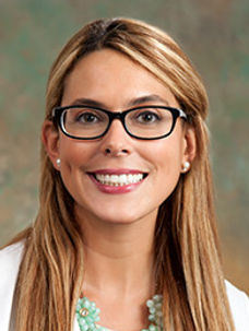
Jaclyn Nunziato, MD
Characterizing the vesicles that are released following high-intensity focused ultrasound (histotripsy) of naturally occurring soft tissue sarcoma
Tumor and circulating extracellular vesicle characterization following mechanical, high-intensity focused ultrasound (histotripsy) in soft tissue sarcoma
Investigators: Shawna Klahn, DVM, DACVIM–Oncology and Natasha Sheybani, PhD
Extracellular vesicles (EVs) are particles naturally released from the cell that are delimited by a lipidbilayer and cannot replicate. EVs transfer genes and protein, acting as two-way communicationlocally and distantly between cells, and are implicated to foster cancer progression and metastasis.EVs also have unique potential to discover new cancer biomarkers, enhance existing treatments, andas a novel drug delivery tool. The impact of high-intensity focused ultrasound (FUS, histotripsy) on vesicle release from cancer cells or the surrounding tissue has never been evaluated, and may have unintended biological consequences, and/or be leveraged as a clinical or research tool.
In this project, Shawna Klahn, an associate professor of Medical Oncology in Virginia Tech, and Natasha Sheybani, an assistant professor of Biomedical Engineering and Research Director of the UVA Focused Ultrasound Cancer Immunotherapy Center in the University of Virginia, characterized the vesicles that are released following histotripsy treatment of naturally occurring soft tissue sarcoma.
Patients with soft tissue sarcoma were treated with histotripsy. Plasma samples were collected before and after the treatment. The researchers developed protocols for shipping and storing these plasmas such that they can be used for analysis of peripheral blood mononuclear cells and EVs. A comprehensive multispectral flow cytometry panel for phenotypic and functional characterization of circulating T cells and myeloid cells have been designed. Multiple EV isolation techniques were developed to confirm EV size distribution and concentration in healthy participants and participants with soft tissue sarcoma individuals. Multiple different panels were tested for characterization of EV surface markers by multispectral flow cytometry and identified promising protein marker candidates to be converged into a single panel. Lastly, the researchers worked up multiple strategies for RNA isolation from EVs, which require further optimization to improve RNA quality. This approach will eventually lend to Nanostring analysis for broad profiling of immune-related gene expression in extracellular RNA payloads.The data generated by this study will advance knowledge and techniques used in the EV-cancer and EV-FUS clinical and research arenas.
Discovery of Novel Probiotic Lactobacillus reuteri Strains to Improve Infant Health
Investigators: Xin Luo, PhD and Monica Garin-Laflam, MD
It was recently discovered in mice that Lactobacillus reuteri, a probiotic bacterium present in the breastmilk, can induce production of immunoglobulin A (IgA), a powerful protective antibody against gut pathogens, in neonatal mice. To translate this knowledge to benefit infant health, Xin Luo, an associate professor of the Department of Biomedical Sciences and Pathobiology in Virginia Tech, and Monica Garin-Laflam, an associate professor of Pediatrics in Virginia Tech-Carilion School of Medicine, tested the hypothesis that infants with higher abundance of specific strains of Lactobacillus reuteri in the fecal microbiome have more IgA capable of recognizing gut pathogens. The identification of neonatal IgA-inducing Lactobacillus reuteri strains will guide formula selection as well as formula design. Future investigations will include the isolation of novel, protective IgA-inducing probiotic Lactobacillus reuteri strains to be used in infant formula, as well as mechanistic studies of how these probiotic strains induce neonatal IgA.
The team successfully enrolled seven healthy, primarily breastfed infants. Infant stool samples at 1, 2,4, 6 months of age were collected. The researchers will analyze these samples in mid-2024 for (1) total fecal IgA; (2) whole-bacteria cross-reactive fecal IgA against gut pathogens (e.g. Salmonella, enterohemorrhagic Escherichia coli (EHEC), and Shigella); and (3) fecal microbiome, specifically of the bacteria bound to IgA, with metagenomic shotgun sequencing.
The discovery of this study will be shared with families with infant children, physicians, and researchers in a facilitated conversation session (Community Engagement Studio).


Shawna Klahn, DVM, DACVIM–Oncology
Natasha Sheybani, PhD

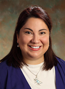
Xin Luo, PhD
Monica Garin-Laflam, MD
The role of altered nutrient partitioning in food reward The role of altered nutrient partitioning in food reward
Investigator: Alexandra DiFeliceantonio, PhD
Obesity is a chronic disease that requires lifelong attention. From a public health perspective, developing long-term effective treatments is an essential medical research goal. The gut-brain axis has long been recognized as an important regulator of metabolism and energy homeostasis; however, only recently have advances in research tools allowed for a better understanding of the complex mechanistic integration of gut and brain signals. Understanding this process presents a novel and potentially powerful therapeutic target for obesity and metabolic diseases.In this proof-of concept pilot project, Alexandra DiFeliceantonio, an assistant professor and associate director at Center for Health Behaviors Research in Virginia Tech, probed the gut-brain axis as a target for obesity intervention. To achieve this, the researcher aimed to:1)determine whether differences in reinforcement learning/flavor-nutrient conditioning of carbohydrate can be measured across the body mass index (BMI) range. Twelve participants across a range of BMI joined this study. The results are consistent with the idea that BMI may influence reinforcement learning. These data provide the first steps in understanding the relationship between overweight/obesity and altered reinforcement learning.

Alexandra DiFeliceantonio, PhD
Uncovering these insights could potentially lead to more targeted drug or behavioral therapeutics for obesity prevention and treatment. 2)determine the feasibility of assessing metabolic flexibility and whether a relationship between metabolic flexibility and calorie-predictive reward can be detected. Participants for part 1 additionally underwent measurements of metabolic flexibility using controlled dietary intake and indirect calorimetry in a metabolic chamber. The goals were to access feasibility and methodologies for assessing metabolic flexibility. The researcher was able to confirm: a) the controlled feeding paradigm established similar between- and within-subjects; 2) the equipment was sensitive enough to capture differing metabolic responses to meals of different macronutrient compositions; and 3) the duration of the measurement sessions, which is much shorter than most other institutions’ protocols, coupled with the small-volume metabolic chamber size, was sufficient to assess metabolic flexibility outcomes. These findings provide important foundational steps for the next step of the research.
A Comparison of the Effectiveness of Telepsychiatry with a Randomized Waitlist Control Utilizing Patient Reported Outcome Measures (PROMs)
Investigators: Anita Kablinger, MD and Lee Cooper, PhD
The utilization of patient reported outcome measures (PROMs) allows for on-going assessment of the severity of mental illness and patient outcomes during treatment. Additionally, it provides feedback on the patient’s psychiatric status to both the patient and practitioner. Carilion Clinic Psychiatry & Behavioral Medicine ambulatory (CC-P&BM) clinic implemented PROMs prior to the start of the COVID-19 pandemic and continues to utilize them as part of patient care. All new patients are asked to complete an initial PROM bundle twenty-four hours before their initial appointment. Research members of Carilion Clinic Psychiatry and Virginia Tech Psychology departments have used PROM data to assess mental health outcomes. Results indicate that patients who received care via telepsychiatry during the pandemic did not experience worsening symptoms, but in fact showed improvements in depression, anxiety and psychological functioning. However, without a control group of untreated patients to compare, the impact of telepsychiatry plus PROMs remains unclear. Researchers Anita Kablinger, a professor in the Department of Psychiatry and Behavioral Medicine at Virginia Tech Carilion School of Medicine, and Lee Cooper, a clinical professor in the Department of Psychology at Virginia Tech, designed a project that randomize waitlist individuals to one of two groups to assess the influence of time while they await initial psychiatric assessment (no intervention) versus minimal intervention using repeated PROMs while awaiting initial psychiatric assessment. This study: 1) Measured the symptomology and well-being of adult individuals on a waitlist group referred to CC-P&BM clinic from their initial referral to their initial psychiatric session with patient-report outcome measures (PROMs) administered monthly (minimal intervention group-MI) versus PROMs administered only at entry to the waitlist and entry to clinic (no intervention group-NI). The researchers found no difference in PROM scores between two groups in depression, anxiety, alcohol/drug use or functional status. Participants who completed a final PROM before their initial appointment were significantly older (38 years vs. 32 years) than those who did not complete a final PROM. Researchers also found longer wait time for the minimal intervention participants. 2) Used PROMs to assess whether there is a difference in clinical symptomology and well-being for patients during tele-treatment with their practitioner compared to waitlist individuals. The researchers were analyzing the results planned to compare the no-intervention group, minimal intervention group, and telepsychiatry group. When the analysis is completed, the findings will be shared.
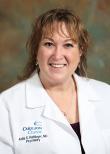
Anita Kablinger, MD
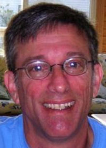
Lee Cooper, PhD
Reducing Operating Room Waste by Monitoring Single-use Sterile Surgical Supplies with Computer Vision
Investigators: Matthew Meyer, MD and Hoda Eldardiry, PhD
Waste has become a hallmark of the US health care—accounting for over 25% of health care expenses. Operating rooms (OR) are major sources of material and financial waste. By one estimate, $2.9 million per year can be saved by not opening single-use, sterile surgical supplies (SUSSS) that were not used. Furthermore, this unused and unnecessarily waste is inevitably disposed of with biohazard waste which needs to either be sterilized or incinerated prior to landfilling—resulting in excessive energy consumption and additional pollution. Systematically identifying unused (wasted) SUSSS at the end of surgery is time intensive and involves handling objects potentially contaminated with blood and human tissue--it is rarely done. Without consistent data assessing wasted SUSSS, it is impossible to quantify the waste and nearly impossible to improve the behaviors that cause the waste. To solve this problem of unnecessary and costly wasted SUSSS in the OR, Matthew Meyer, an associate professor of Anesthesiology in the University of Virginia Health, and Hoda Eldardiry, an associate professor in the Department of Computer Science at Virginia Tech, aimed to develop computer vision software that identifies unused SUSSS on the scrub table at the end of surgery.
The researchers: 1) Analyzed pilot data from pediatric ORs to quantify wasted SUSSS in an analog manner. After completing the analyses, the findings were shared locally and nationally with health care providers. 2) Designed a computer vision software for SUSSS identification and analysis. A proof-of-concept software was developed. It is a computer-vision assistance with hand-tracking to highlight the moments when items are manipulated and may be used. This software allowed perioperative administrators to visualize the impact of computer vision on reviewing intraoperative sterile surgical item usage.

Matthew Meyer, MD
Hoda Eldardiry, PhD
Advanced Immunoclinical phenotyping of rejection in lung transplant Investigators: Y. Michael Shim, MD and Jaime Mata, PhD
Lung transplantation is a therapy for many patients with end-stage lung diseases. Unfortunately, studies report that most transplant recipients die from rejection. Rejection occurs because the lung transplant recipient’s body perceives the donor’s lungs as foreign and rejects the transplanted lungs. Many now believe that a series of small rejections lead to terminal failure of the transplanted lungs. The supply of lungs for transplant is the biggest bottleneck to perform a lung transplant, and many patients die while waiting. Therefore, it is critical to detect rejection early and treat it aggressively so that already-transplanted lungs can give the longest possible survival benefit to the recipients. Some lung transplant centers (like UVA Health) perform routine invasive lung sampling procedures called a bronchoscopy, pulmonary function test, and clinical exam to diagnose rejection. Lung sampling is typically performed in the right middle or left upper part of the lung because those areas are easy and convenient to sample. Unfortunately, many patients develop rejection unpredictably, after lung samplings showing normal results. The failure to effectively diagnose the rejection by bronchoscopy raises questions. Are clinicians sampling correct parts of the lung to diagnose rejection, and can we improve our lung sampling techniques?
A specialized MRI technique called hyperpolarized gas MRI (HGMRI) has been used to visualize and detect specific parts of the lung with abnormalities. Researchers, Michael Shim, an associate professor of medicine in the Division of Pulmonary and Critical Care Medicine at the University of Virginia, and Jamie Mata, a research professor of Radiology and Medical Imaging in the University of Virginia, showed that the rejection happens in moth-eaten-like random patterns with normal and abnormal lung tissues alternating. These results suggest that the current way of sampling the right middle or left upper lung by bronchoscopy may be severely flawed.
In this pilot project, the researchers studied lung transplant patients who were scheduled to undergo surveillance bronchoscopy to detect rejection. The researchers first found the part of the patients' lung with rejection-like changes by HGMRI, then sampled the lung in two places, one normal and one abnormal area of the lung. These two areas of the lung from the same patient were compared to determine if abnormal findings in HGMRI correlate with rejection.


Y. Michael Shim, MD
Jaime Mata, PhD
In part 1 of this pilot project, 12 patients were imaged using HGMRI. As hypothesized, HGMRI was able to differentiate focal regions in the transplanted lungs that are abnormal and not performing well. The researchers developed an imaging tool that fuses the HGMRI images with the CT scan and produces a detailed 3D “path map” for the bronchoscopy, from the trachea all the way to the focal lung abnormality. With this imaging analysis tool, the researchers can provide 3D guidance ahead of the bronchoscopy procedure, helping in a novel and unparalleled way the planning of the bronchoscopy procedure.In part 2 of the project, with the new imaging tools developed for this project and described above, the researchers were able to sample the abnormally functioning regions of the allografts (transplanted lung) with an unmatched planning and precision. The researchers also have been able to determine normal functioning regions of the allografts for comparison. The preliminary results have been astonishing, and in several participants, the researchers were able to correlate initial allograft rejection from the standard clinical pathology samples with the abnormal HGMRI ventilation and gas exchange imaging.This new pre-bronchoscopy personalized planning developed for this project is having a tremendous impact in the way the physicians at the University of Virginia perform their bronchoscopies. It is also helping the UVA patients in getting a more precise, an earlier diagnosis and earlier modified treatment plan in case of abnormal lung function or signs of early lung rejection.
Neural circuit mechanisms for decoding self-harm
Investigators: Sora Shin, PhD
Self-harm refers to the intentional act of physical damage to one’s own body. It is particularly prevalent among teenagers and young adults and is often accompanied by an elevated risk of suicide. The risk assessment of self-harm is extremely difficult to determine due to reliance on self-reports, which is limited by an individual’s lack of insight and desire to conceal such intentions. The identification of objective information gained from neurobiological underpinnings of self-harm is a critical next step for the development of a novel prevention/therapeutic strategy for the self-destructive behaviors in numerous psychiatric disorders.
Researcher Sora Shin, an assistant professor in the Fralin Biomedical Research Institute at Virginia Tech Carilion, set out to 1) examine the circuit-specific role of the thalamic nucleus reuniens (RE) region of the brain in controlling self-harm

Sora Shin, PhD
and its neural adaptation following early adversity, and 2) determine how early life trauma remodels molecular composition in the cell-type specific RE neurons, given that early life trauma is a critical risk factor for self-harm in later life.Using genetic, molecular, and neurophysiology tools, the researcher identified neurons expressing the gene vesicular glutamate transporter (vGlut2) in the thalamic nucleus reuniens (RE) that are projected to the ventral hippocampus (vGlut2 RE->Hip) as the neural circuit mechanisms underlying self-harm. Furthermore, it was found that early life trauma -induced molecular changes (upregulated L-type calcium channels in the thalamic nucleus reuniens) are associated with increased vulnerability to self-harm. In conclusion, the results identified circuit-based targets and promising candidates for pharmacological intervention in self-harm.
Sora Shin, PhD
Defining donor characteristics for pediatric heart transplants
Impact Quantification of Donor Echocardiographic Data on Pediatric Heart Transplantation Recipient Outcomes
Investigators: Michael McCulloch, MD and Michael Porter, PhD
Heart transplantation is the standard of care for pediatric patients with end-stage heart failure or inoperable congenital defects, yet nearly 20 percent of patients with these conditions die while on the waitlist. To help increase the odds of successful pediatric heart transplants, Michael McCulloch, an associate professor and a pediatric cardiologist at UVA Children’s Hospital Heart Center, and Michael Porter, an associate professor of systems engineering in UVA’s School of Engineering and Applied Science, set out to evaluate the entirety of the US experience in how pediatric donor heart offers were being utilized when offered to pediatric heart transplant candidates.
The literature has always suggested a national shortage of pediatric heart donors, with transplant institutions willing to be aggressive to accept ‘marginal donors’ (e.g. decreased heart function, requiring high volume inotrope support). This interdisciplinary team of researchers, consists of pediatric transplant cardiologists, data scientists, undergraduate students and systems engineering students, collected and cleaned large amount of heart transplant data, analyzed millions of pages of documents to compile the most complete representation of pediatric donor heart function measurements ever collected. These measurements have been inadequately assessed but universally recognized as crucial in the decision-making process of accepting / rejecting a donor's heart. The researchers found that the overarching problem with the way this process was handled could be attributed to behavioral economics as evidenced by the following:
-
Less than 10% of all offers accepted had any abnormality of heart function, despite the stated willingness to accept marginal hearts for the sickest patients
-
90% of all offers were refused and 40% of pediatric donor hearts were NEVER utilized by a pediatric institution (despite having been offered to them). However, more than half of these hearts not utilized for pediatric waitlisted patients were utilized by adult institutions or pediatric heart transplant institutions in Canada
-
The most important variable influencing heart offer acceptance was how many prior institutions had already declined it
-
A pediatric transplant institution’s prior year practice/outcomes was the second most important variable determining whether a heart offer was accepted.
These findings have led to follow-on funding, enabling researchers to develop predictive mathematical models to assist transplant institutions in the decision-making process of accepting/rejecting pediatric donor's hearts.

Michael McCulloch, MD

Michael Porter, PhD
Designing an interactive training system for pediatric telemedicine cart operations incorporating augmented reality
Investigators: Elham Morshedzadeh, PhD and Lydia Hartzell, MPH
Elham Morshedzadeh, an assistant professor of industrial design in Virginia Tech’s College of Architecture and Urban Studies; Andre Muelenaer, a professor of practice in Virginia Tech’s College of Engineering, and Carilion’s Lydia Hartzell, designed a robust and affordable training program to help improve telemedicine encounters for infants and pre-school children. They sought to provide a robust, feasible, and affordable training program integrating augmented reality, online and hands-on learning experience.
The team's research focused on how Augmented Reality (AR) can be employed in the healthcare training system to increase efficiency in Lean Healthcare (LH) and to improve telemedical visits for infants and children. They employed Telemedicine Carts which entails systems that integrate cameras, displays, and network access to bring remote physicians right to the side of the patient. This allows patients to communicate with a healthcare provider using technology, as opposed to physically visiting a doctor's office or hospital. To achieve this, the researchers proposed a training program via augmented reality, which will improve both online and in-person learning experiences.
AR represents digital information on top of real-world environments and in this study, it is utilized as interactive learning support to increase engagement and immersion in tele-healthcare. Digital and virtual objects (e.g., graphics, text, sounds) are superimposed on an existing environment to create an AR learning experience for telemedicine operators. The training system has been designed on the hints received from both contextual inquiry (Qualitative) and eye-tracking (Quantitative) techniques to inform the design solutions. The investigators used various tools for data collection from qualitative methods such as contextual inquiry, observation to a quantitative method, eye tracking to ensure triangulating the data. Combining eye-tracking techniques with other research techniques such as observation and contextual inquiry led to a holistic understanding of users’ needs and opportunities associated with AR training systems in telehealth care.
The researchers' next steps will focus on an AR Prototype and its immersive and intuitive experiences that benefit the four parties involved in this research (patients, parents, doctors, and nurses/operators).

Elham Morshedzadeh, PhD

Lydia Hartzell, MD
Measuring medication in patients with epilepsy
Effect of various neuroactive drugs on frequency distribution and phase coupling in various human brain regions- Neuroactive drugs and lntracranial EEG (iEEG) study
Investigators: Aashit Shah, MD and Sujith Vijayan, PhD
Aashit Shah, and a professor of internal medicine at the Virginia Tech Carilion School of Medicine, and Virginia Tech’s Sujith Vijayan, an assistant professor in the Virginia Tech College of Science’s School of Neuroscience studied patients with intractable epilepsy who have been implanted with electrodes to determine the region responsible for their seizures. The team examined signals measured in epileptic patients undergoing intracranial electroencephalography. They reviewed intracranial electrical signals from various brain regions following administration of medications that work on the brain.
The investigators have thus far focused their efforts on the limbic system and frontal cortices. These structures are associated with emotional experiences and memories and are thought to be important in processing pain.
The team have observed the following:
Fentanyl and Hydrocodone, both opioids, impact spectral characteristics similarly, yet also have distinctive features as seen in their differential effects on limbic structures and frontal cortices. For example, in the left amygdala, a limbic structure, both drugs suppress lower frequency activity, but at the same time, fentanyl also increases higher frequency activity. In contrast, in the right superior frontal cortex, both drugs suppress lower frequency activity, but the exact frequencies that are suppressed within in this range are different from those suppressed in the amygdala, for both drugs. Furthermore, fentanyl does not increase higher frequency activity in the right superior frontal cortex.
The common features of the actions of these drugs in limbic and frontal regions could help to identify the invariant manners in which opioids act as a class of drugs. At the same time, their distinguishing features could help to pinpoint the mechanisms through which particular opioids affect the brain in different ways.
The investigators plan to continue analyzing these data. One area of focus will be an examination of the effects of all opioids versus a comparison drug commonly administered to patients during their stay, such as an antibiotic or anti-nausea medications. The investigators will also compare and contrast how opioids individually compare to one another as well as collectively and individually versus the effects of Fioricet (acetaminophen + butalbital + caffeine), a non-opioid pain medication. The team will also examine how the above comparisons are impacted by gender and prior history of addiction.
.jpg)
Aashit Shah, MD
Sujith Vijayan, PhD
Searching for genetic markers for celiac disease with machine learning
Use of machine learning image analysis and tissue transcriptomics to define clinically actionable celiac disease sub-types
Investigators: Sana Syed, MD and Suchitra Hourigan, MD
Sana Syed, an assistant professor in the UVA School of Medicine’s department of pediatrics, and Suchitra Hourigan, Inova Children’s Hospital’s vice chair of research and innovation sought to determine if machine learning is useful in diagnosing celiac disease sub-types. Currently, treatment and management of celiac disease involves gluten-free diet and is not intended to help assess the specific for the risk of patients developing other diseases. Syed and Hourigan investigated gut tissue biopsies as well as genetic markers of patients with celiac disease and type 1 diabetes and/or hypothyroidism.
In Aim 1, the investigators sought to leverage an existing machine learning small bowel image analysis platform to distinguish & predict celiac disease (CD) sub-types. They hypothesized that their model would identify a spectrum of known features (inflammatory cells, intestinal epithelial morphology) and novel features related to inflammation, tissue injury, and epithelial regeneration that will distinguish and enable the prediction of CD sub-types. The team’s existing CD image analysis platform was trained using duodenal biopsies at the time of diagnosis of CD without Type 1 Diabetes or hypothyroidism versus CD with either condition. Biopsy data originated from CD patients at INOVA and UVA. On visualization using saliency maps, they found distinctive histopathologic features detected by the model, including inflammatory cells, the morphology of intestinal epithelium, and entero-endocrine cells. The investigators scanned and digitized slides from all patients enrolled at both sites and labeled them using the Marsh scoring system. Two pathologists at UVA did this with a tie-breaker pathologist from an external site. The investigators then trained their machine learning model to differentiate between celiac disease and normal and Marsh scores 1-3. The team found distinctive differences in saliency maps between early and severe diseases.
Aim 2 and Aim 3 sought to explore the gene expression profiles for CD sub-types using Formalin-Fixed Paraffin-Embedded (FFPE) duodenal biopsy samples. Using archival FFPE samples for each CD sub-type, the investigators planned to extract and isolate RNA and sequence it to obtain differentially expressed genes. After consenting their enrolled population of transcriptomic sequencing, the team contacted an external vendor, GENEWIZ, who provided quotes for generating high-quality RNAseq. All patients diagnosed post-2015 were consented according to NIH guidelines. The investigators are currently in the process of shipping tissue blocks of these patients to GENEWIZ, and will use discretionary funds to complete Aims 2 and 3.
Sana Syed, PhD
Suchitra Hourigan, MD
Studying auditory therapy for Parkinson’s disease
Sound Sleep with Parkinson's: Nighttime Auditory Therapies to Improve Learning and Impede Disease Progression
Investigators: Della Williams, MD and Sujith Vijayan, PhD
Della Williams, a neurologist at Carilion Clinic and an assistant professor of internal medicine at the Virginia Tech Carilion School of Medicine partnered with Sujith Vijayan, to study auditory therapy for patients living with Parkinson’s disease. Their project sought to better understand the disease progression and if it can be slowed by providing some background noise to patients as they sleep.
In order to better pinpoint PD-related aberrant brain dynamics and develop non-invasive, innocuous interventions during sleep that improve motor learning in PD and potentially impede the progression of the disease itself, the following aims were pursued:
Aim 1: To strengthen the evidence for a linkage between aberrant sleep spindles in slow wave sleep and PD.
Aim 2: To evaluate the effect of auditory intervention during sleep on motor learning in PD patients.
The data collected so far indicate the following:
PD patients do not show overnight improvement in their reaction time in a finger tapping task.
PD patients spend a smaller percentage of their time in stage 3 non-rapid eye movement sleep (N3 sleep) than do control subjects.
There is less power in the slow wave frequency band in the frontal and central channels during N3 sleep in PD patients in comparison with control subjects.
There is less power in the spindling band in the frontal and central channels during N3 sleep in PD patients in comparison with control subjects.
There are indications of aberrant spindle-slow-wave coupling in PD patients.
In the coming months, the investigators plan to collect additional data to solidify these initial findings. The team also plans in the next step to see whether they can restore normal sleep dynamics in PD patients using auditory stimuli during sleep and improve their learning of motor tasks (e.g., the finger tapping task)—which would clearly have great clinical implications.
Once they have collected additional data, the team will apply for additional grant funding to support this line of research.

Della Williams, MD

Sujith Vijayan, PhD
When Can Patients Safely Drive After Rotator Cuff Repair?
Investigators: Peter Apel, MD and Miguel Perez, PhD
Despite more than 450,000 rotator cuff repairs performed every year, very little data exists guiding when a person can return to driving. Dr. Peter Apel, an assistant professor at Virginia Tech Carilion School of Medicine and a Carilion Clinic orthopedic surgeon, and Miguel Perez, an associate professor at the Virginia Tech Transportation Institute, lead this team. The team investigated how changes in driving skills can shorten the time patients are restricted from driving after surgery.
The researchers were able to determine that the driving fitness of patients who had undergone rotator cuff repair surgery (RTCR) was non-inferior to a baseline preoperative reading. Specifically, patients showed no clinically significant negative impact on driving fitness as early as two weeks after RTCR. This has significant clinical implications, as it is currently common practice for physicians to restrict patient driving after RTCR until as far as six to eight weeks after surgery.
Next, the researchers plan (a) to publish their conclusions immediately in a major orthopaedic journal to disseminate these findings to the surgeons who routinely perform this procedure and (b) to utilize what they learned to investigate other forms of postoperative driving restrictions in orthopaedic surgery, such as after total knee arthroplasty.

Peter Apel, MD

Miguel Perez, PhD
Video courtesy of Carilion Clinic
